

African horse sickness (AHS) is a viral disease affecting equids, with mortality rates reaching 90% in naive horse populations (King et al, 2020). The causative agent, African horse sickness virus (AHSV), belongs to the genus Orbivirus within the family Sedoreoviridae (Weyer, 2022). AHS was confirmed in horses in Thailand during March 2020. The virus belongs to serotype 1 and is phylogenetically closely related to isolates from South Africa. This represents the first incidence of AHS in Southeast Asia and the first detection of serotype 1 outside Africa (King et al., 2020).
Regarding agent identification, it is particularly important to perform virus isolation and serotyping whenever AHS outbreaks occur outside enzootic regions to ensure the selection of a homologous vaccine serotype. AHSV can be isolated from whole blood collected during the early febrile stage (WOAH, 2019). Post-mortem tissue samples used for virus isolation typically include the spleen, lung, and lymph nodes collected during necropsy (WOAH, 2019; Zeleke, 2005).
For virus isolation, sample preparations can be inoculated into mammalian cell lines—such as baby hamster kidney (BHK-21), monkey stable (MS), and African green monkey kidney (Vero)—as well as Culicoides or mosquito insect cell lines. Alternatively, samples may be inoculated intravenously into embryonated chicken eggs (WOAH, 2019).
Virus isolation was performed using BHK-21 (CLS C-13), Vero (ATCC CCL-81), and Aedes albopictus clone C6/36 (ATCC CRL-1660) cell lines. Undiluted blood samples, collected in EDTA from horse farms in Thailand in March 2020, served as the inoculum after testing positive for AHS by RT-PCR.
Confluent Vero, BHK-21, and Aedes albopictus clone C6/36 cells seeded in 12-well plates were inoculated with 100 µL of each blood sample. The Vero and BHK-21 plates were incubated at 37°C with 5% CO₂, while the C6/36 plates were incubated at 27°C with 3% CO₂ for 3–4 days. Upon observation of cytopathic effects (CPEs) or at the end of the incubation period, supernatants were harvested for blind passage. The harvested supernatants were then inoculated into newly prepared cell monolayers for a second passage. This blind passage protocol was continued for up to five passages in Vero cells (Figure 1) and BHK-21 cells (Figure 2), and four passages in C6/36 cells. Finally, the viral supernatants were harvested and analyzed via RT-PCR for diagnostic confirmation. The test results confirmed that the C6/36 cells were positive for AHSV.
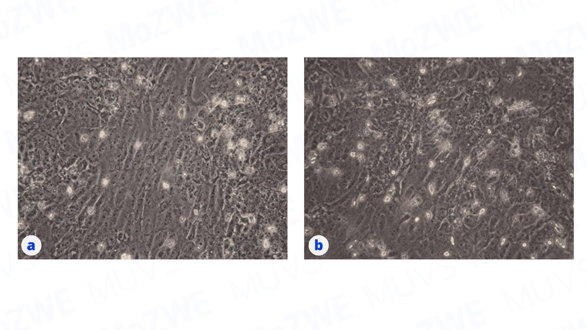
Figure 1 Morphological appearance of Vero cells under an inverted microscope.
(a) Vero cell control (10×). (b) AHSV-infected Vero cells at passage 5 (20×).
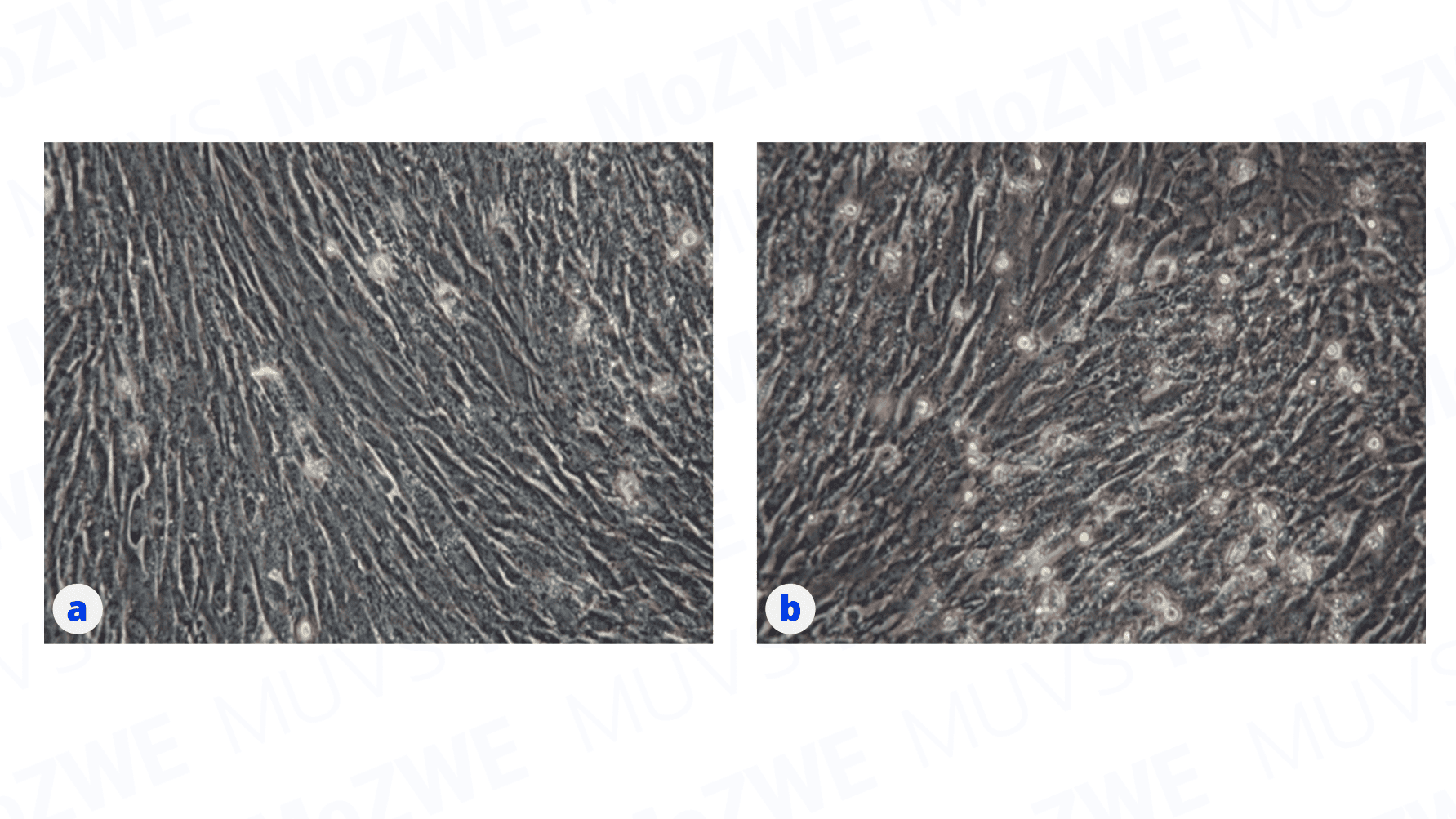
Figure 2 Morphological appearance of BHK-21 cells under an inverted microscope.
(a) BHK-21 cell control (10×). (b) AHSV-infected BHK-21 cells at passage 5 (20×).
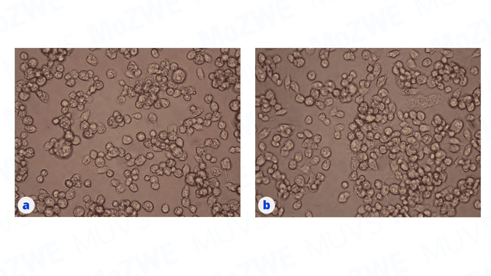
Figure 3 Morphological appearance of Aedes albopictus clone C6/36 cells under an inverted microscope.
(a) Aedes albopictus clone C6/36 cell control (10×). (b) AHSV-infected Aedes albopictus clone C6/36 cells at passage 4 (20×).
Although AHSV isolation in Aedes albopictus clone C6/36 cells did not produce any CPE (Figure 3), the virus remained active. We harvested the supernatant from C6/36 passage 4 and inoculated it into new Vero and BHK-21 cells. While continuing cultivation for 5 passages, we observed clear CPEs in BHK-21 cells (Figure 5) but not in Vero cells (Figure 4). The complete results for AHSV isolation in cell culture are summarized in Table 1.
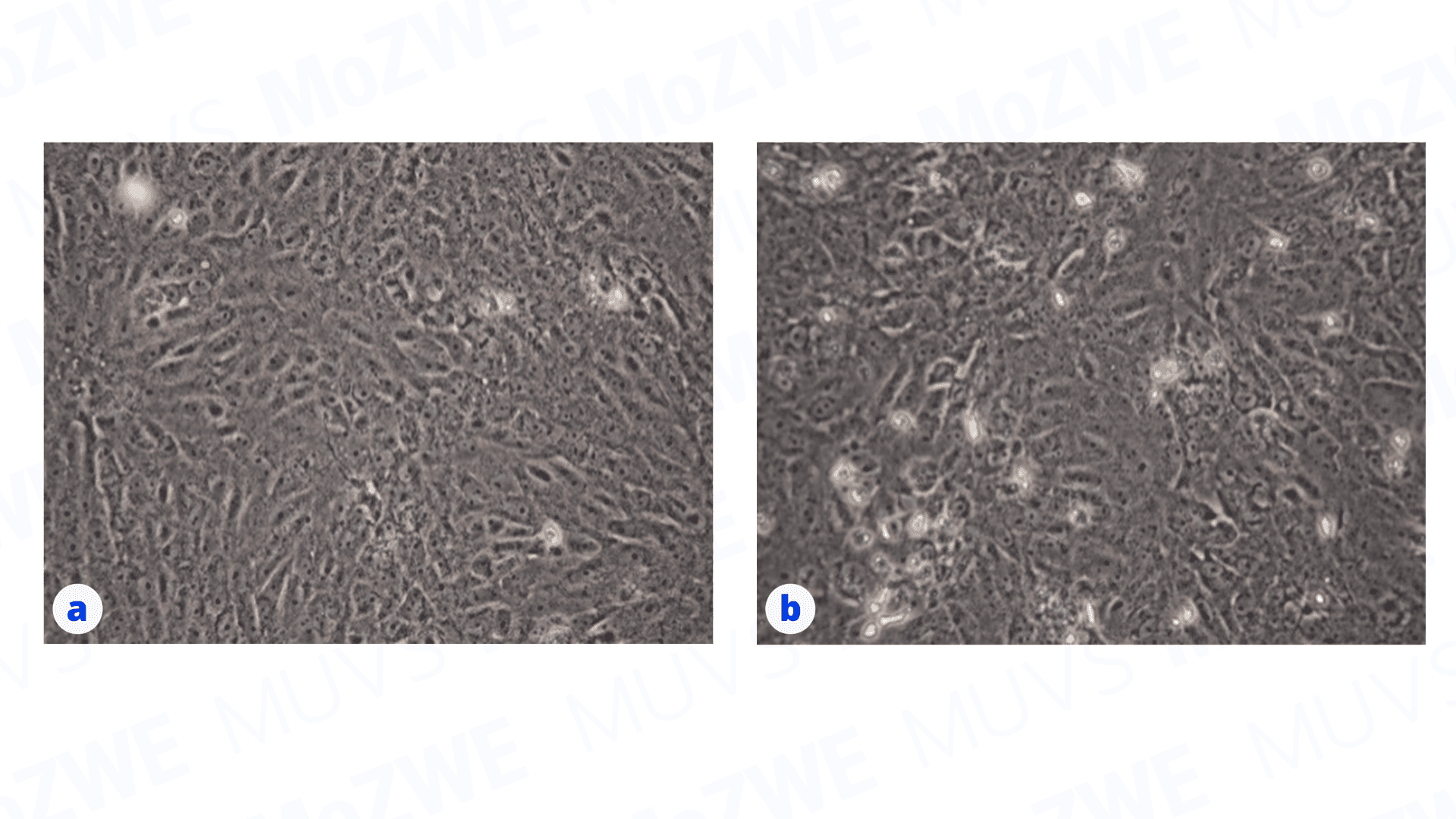
Figure 4 Morphological appearance of Vero cells under an inverted microscope.
(a) Vero cell control (20×). (b) Vero cells inoculated with passage 4 supernatant from C6/36 cells, imaged at passage 5 (20×).
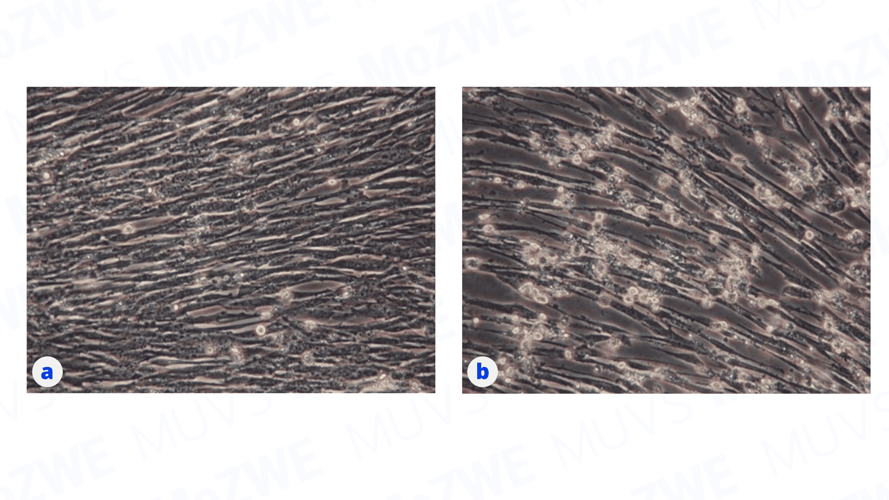
Figure 5 Morphological appearance of BHK-21 cells under an inverted microscope.
(a) BHK-21 cell control (20×). (b) BHK-21 cells inoculated with passage 4 supernatant from C6/36 cells, imaged at passage 5 (20×).
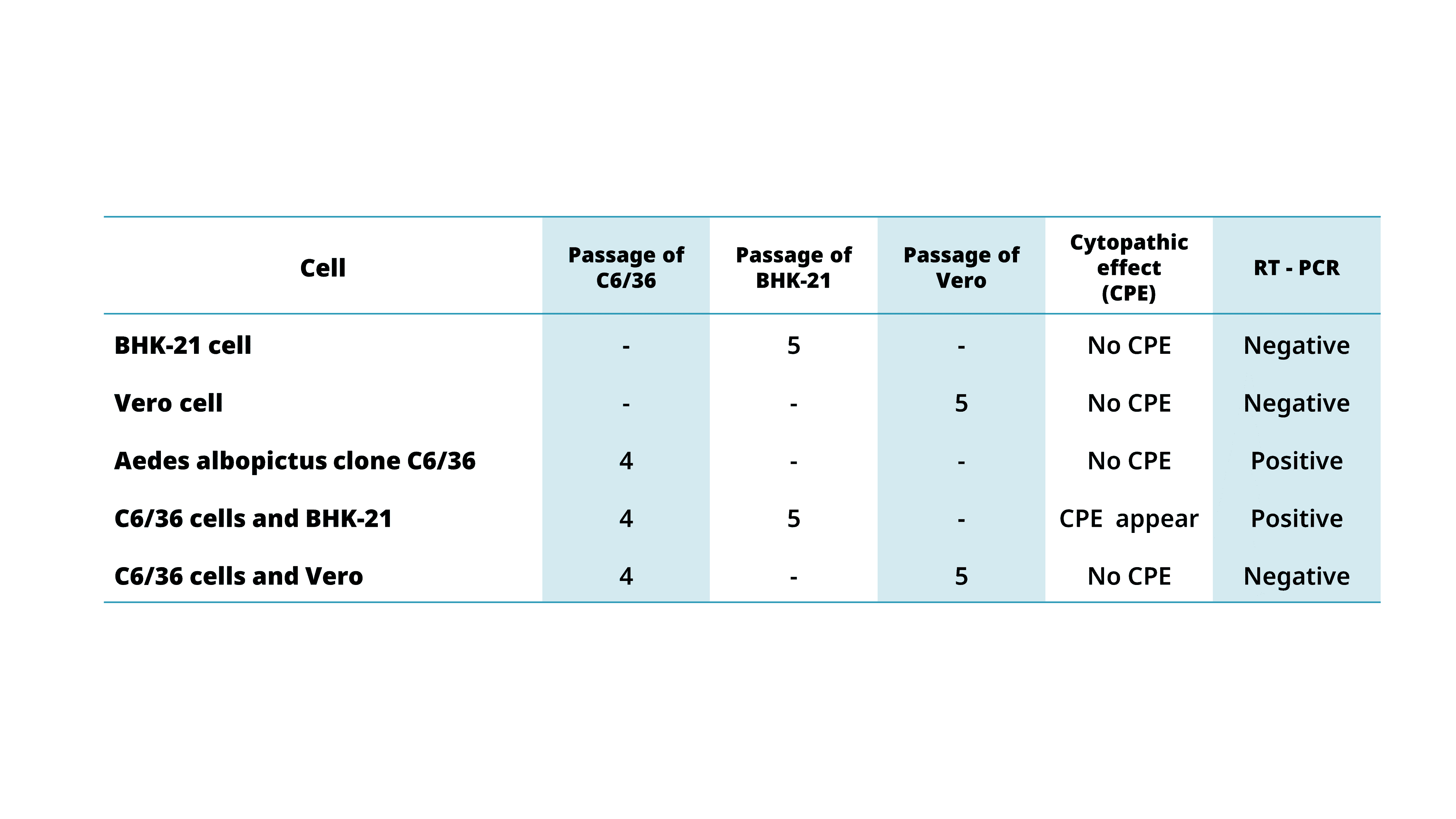
Table 1 Summary of the results of AHSV isolation in cell culture
To optimize AHSV isolation, selecting an appropriate host cell line is critical for maximizing diagnostic sensitivity. Furthermore, sample integrity depends heavily on temperature management; specimens must be maintained at 2–8°C and transported to the laboratory immediately. Maintaining a cold chain throughout sample preparation and ensuring rapid host inoculation are vital. Samples intended for live virus isolation should not be frozen, as freeze-thaw cycles can significantly reduce viral viability.
Reference
King S, Rajko-Nenow P, Ashby M, Frost L, Carpenter S, Batten C. Outbreak of African horse sickness in Thailand 2020. Transboundary and emerging diseases 2020; 67: Pages i-viii, 1751-2267. https://onlinelibrary.wiley.com/doi/epdf/10.1111/tbed.13701
Zeleke A, Sori T, Powel K, Gerbre-Ab F, Endebu B. Isolation and Identification of Circulating Serotypes of African Horse Sickness Virus in Ethiopia. The journal of applied research in veterinary medicine 2005; 3: 41-3.
Weyer C. African Horse Sickness – Generalized Conditions. MSD manual veterinary manual professional version. 2022. https://www.msdvetmanual.com/generalized-conditions/african-horse-sickness/african-horse-sickness#
World Organisation for Animal Health (WOAH). African horse sickness (infection with African horse sickness virus). WOAH Terrestrial Manual 2019; Chapter 3.6.1: 1-17.

Jarupha Taowan
Scientist (Senior Professional Level), The Monitoring and Surveillance Center for Zoonotic Diseases in Wildlife and Exotic Animals (MoZWE)